

Specialist care
Leg vein and artery ultrasound in Slavuta
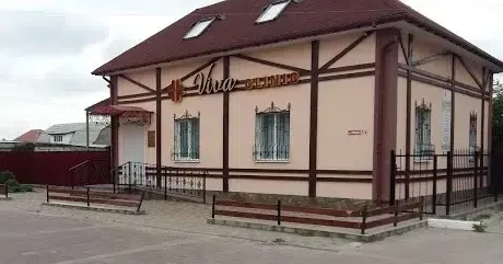
A color Doppler ultrasound of the leg vessels, also called a duplex scan, shows whether the vein valves work, whether there are blood clots and whether the arteries are narrowed. At Viva Clinic in Slavuta it is done at the polyclinic at 27a Sobornosti St, and varicose vein treatment starts with this scan. You can book by phone or on Viber.
Paid serviceBy appointment
At a glance
- Where
- 27a Sobornosti St, Slavuta
- Hours
- Mon–Fri 8:00–17:00
- How to book
- phone or Viber
- Payment
- paid
When you may need a leg vessel ultrasound
- Heavy, tired or swollen legs at the end of the day
- Spider veins or enlarged veins under the skin
- Night cramps in the calves
- Darkened or hardened skin on the lower legs
- Calf pain when walking that goes away after a short rest
- Cold, pale feet or sores on the legs that are slow to heal
- Preparing for varicose vein treatment, including laser treatment
- Varicose veins in the family, if you want your veins checked before symptoms appear
What a leg vein and artery ultrasound includes
- Color Doppler duplex scan of the leg veins
- Ultrasound of the leg arteries
- Checking the vein valves for backward blood flow (reflux)
- Scanning the deep and superficial veins to rule out a blood clot
- Vein ultrasound before choosing varicose vein treatment, including before EVLA
- An ultrasound report for your vascular surgeon or family doctor
- A consultation with a vascular surgeon (phlebologist) at the clinic
What a leg vessel ultrasound shows
A standard ultrasound shows the vessel walls and the space inside. Color Doppler adds the movement of blood, so the doctor can see which way it flows and how fast. Combining the two modes is what’s called a duplex scan.
In the veins, the doctor checks the valves. Healthy valves let blood flow only upward, toward the heart. If a valve doesn’t close, blood flows back down, which is called reflux. Twisted veins under the skin often appear because of a problem in a large vein that can’t be seen from the outside, so without an ultrasound the doctor can’t choose a treatment. The doctor also checks the deep and superficial veins for blood clots.
In the arteries, the scan shows plaques, narrowed sections and places where blood flow is blocked. Arteries are checked when your calves hurt when you walk, your feet are cold or sores on your legs heal poorly.
What happens during a leg vein and artery ultrasound
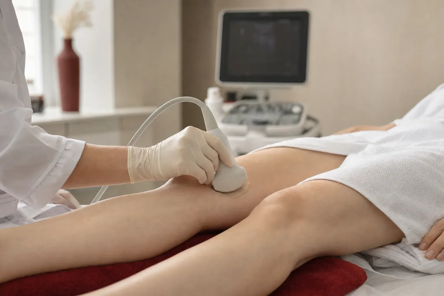
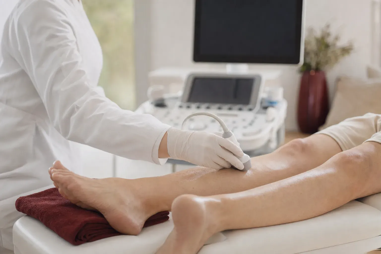
No special preparation is needed. You’ll be asked to undress to your underwear. Veins are mostly checked while you stand, because then the weight of the blood presses on the valves and shows whether they work. The doctor moves the probe with gel from the groin to the foot, squeezes your calf by hand or asks you to hold your breath and bear down, and watches where the blood flows. The doctor also checks the deep veins by pressing on them with the probe. Arteries are usually scanned while you lie on your back.
You will feel the pressure of the probe, and it may be uncomfortable over a sore vein. If you have had a vein ultrasound before, bring the report so the doctor can compare the results.
What happens after the ultrasound
The ultrasound report is reviewed by a Viva Clinic vascular surgeon. If you book directly with the surgeon, they will do the color Doppler vein scan themselves after examining your legs. The doctor then compares the exam with the results and suggests a treatment plan. Laser treatment doesn’t suit every vein: for example, a vein may be too twisted or lie too close under the skin. So the vascular surgeon chooses the treatment method, including whether laser treatment of varicose veins (EVLA) suits you, based on the ultrasound results. The article on early signs of varicose veins describes how the condition usually starts, and the article on laser varicose vein treatment explains the procedure and recovery.
It is also better to buy compression stockings after an exam and ultrasound. Compression should not be used with some diseases of the leg arteries, and poorly fitted stockings squeeze the leg and hinder blood flow. The doctor will advise on the class and size.
A leg vessel ultrasound at Viva Clinic is paid. Patients come to Slavuta for varicose vein tests and treatment from Netishyn, Shepetivka, Khmelnytskyi, the Khmelnytskyi and Rivne regions and from all over Ukraine. Other tests at the polyclinic are listed on the ultrasound and diagnostics page.
When you need urgent help
See a doctor the same day if one leg suddenly swells and hurts, especially in the calf, or a vein becomes hard, red and painful. These can be signs of a blood clot.
Call an ambulance on 103 if you suddenly become short of breath, have chest pain or cough up blood. You also need an ambulance if a leg suddenly turns cold and pale, goes numb and hurts badly, or if a varicose vein bleeds. Until the ambulance arrives, lie down, raise the leg and press firmly on the wound with a clean cloth.
Questions about leg vessel ultrasound
How do I prepare for a leg vein ultrasound?
No special preparation is needed, and you can eat and drink as usual. Wear clothes that are easy to take off down to your underwear, because the doctor scans the leg from the groin to the foot. If you have had a vein scan or varicose vein treatment before, bring the reports.
Why is a vein ultrasound done standing up?
When you stand, the weight of the blood presses on the vein valves, which shows whether they close tightly. Lying down, backward flow may not show up. That is why European vascular surgery guidelines (ESVS) recommend checking for reflux standing, or sitting with the legs hanging down if a person cannot stand.
Can an ultrasound show a blood clot in a vein?
Yes. A clot shows on the screen, and the doctor also presses on the vein with the probe: a healthy vein squeezes flat, one with a clot does not. If one leg suddenly swells and hurts, see a doctor the same day. If you also feel short of breath or have chest pain, call an ambulance on 103.
Do I need a vein ultrasound if I only have spider veins?
It is a good idea. Spider veins are often just a cosmetic issue, but sometimes they come with hidden problems in deeper veins. The ultrasound shows how the valves work, and the vascular surgeon decides whether you need treatment and which kind.
How is an artery ultrasound different from a vein ultrasound?
In veins the doctor checks the valves and looks for clots; in arteries the doctor looks for narrowing and plaques and checks how blood reaches the foot. Arteries are usually scanned lying down. The doctor decides what to check based on your symptoms: heaviness and swelling usually point to the veins, calf pain when walking to the arteries.
Articles: leg vein and artery ultrasound
Varicose veins: what they are, early signs and treatment
Varicose veins are enlarged leg veins that develop when the valves inside them weaken and blood pools. The first signs are usually heavy, swollen legs in the evening, spider veins and night cramps, and later twisted veins become visible under the skin. Treatment starts with a vein ultrasound, and based on the results a vascular surgeon chooses compression, a minimally invasive procedure or surgery.
When to have a breast ultrasound and on which day of your cycle
A routine breast ultrasound is usually done in the first half of your cycle, roughly between days 5 and 12 counting from the start of your period, when your breasts are least swollen. After menopause, any day is fine. If you've noticed a lump or nipple discharge, don't wait for the right day and book an appointment with a mammologist, a breast specialist, straight away.

Laser treatment for varicose veins: preparation, procedure and recovery
Laser treatment for varicose veins, or endovenous laser ablation (EVLA), closes the affected vein from the inside through a small puncture, with no incisions. It is done on an outpatient basis, and a vascular surgeon decides whether it suits you after an exam and a vein ultrasound. After EVLA you usually go home the same day, and the doctor tells you what activity level and compression stockings you need.

What kidney pain feels like and how to tell it from back pain
Kidney pain is felt in the side of your lower back, just under the lowest ribs, usually on one side. It is dull and aching or comes in waves, as with renal colic, hardly changes when you move and often comes with a fever or changes in your urine. Back pain usually gets worse when you bend, twist or sit for a long time and eases when you rest.

Ligament sprain: what to do and when to see a trauma doctor
In a sprain, some of the fibers in a ligament tear, so the joint hurts and swells, a bruise appears and moving it is painful. For the first 48 hours, rest the joint, apply cold through a cloth, wrap it in a moderately firm bandage and keep your leg raised. If you cannot stand on your leg or the joint looks deformed, go to a trauma unit (travmpunkt), where they will take an X-ray straight away. If the pain does not ease within a few days, see a trauma doctor to rule out a fracture or a torn ligament.
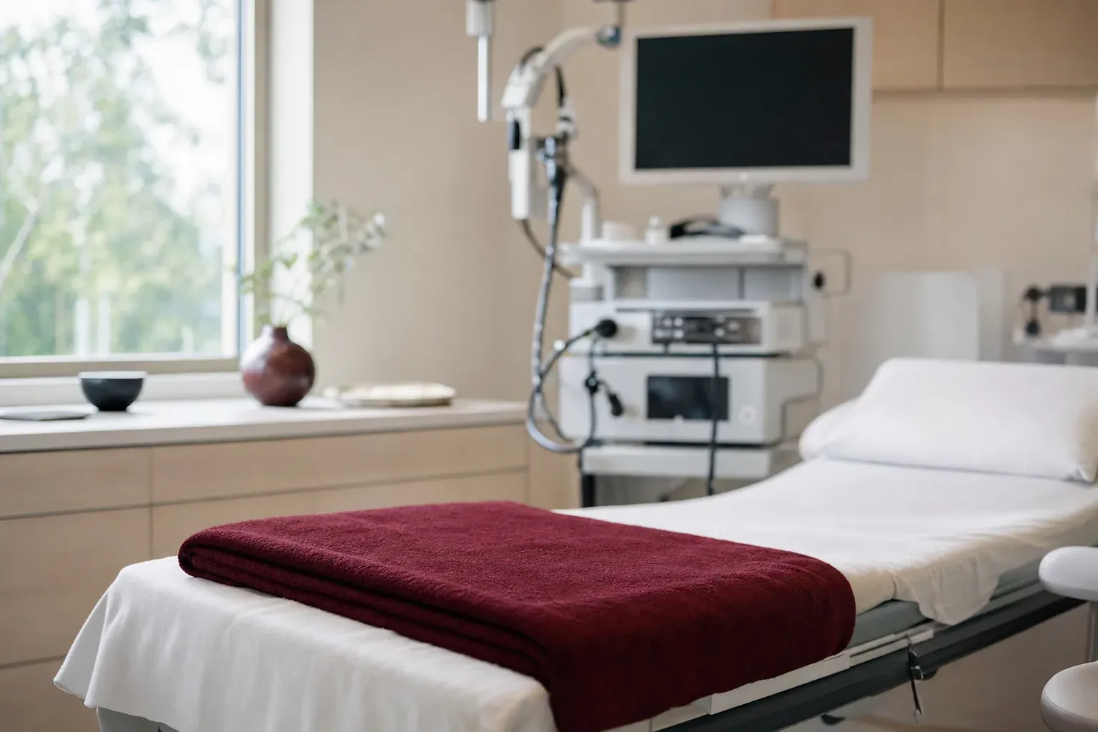
Colonoscopy: what it is, how to prepare and whether it hurts
During a colonoscopy, a doctor examines the inside of the large intestine with a thin, flexible tube that has a camera on the end. So that the doctor can see the whole lining, the bowel is cleared in advance: a few days before, you switch to low-fiber food, and the day before, you drink a bowel prep solution as your doctor instructs. Without anesthesia the test can be unpleasant, so you can have it under anesthesia.

How to prepare for an abdominal ultrasound
Come to an abdominal ultrasound on an empty stomach, usually after 6–8 hours without food, which is why the scan is easiest to schedule in the morning. You can drink a little plain water. If you often get bloated, cut out the foods that make it worse for 1–2 days before the scan.

Thyroid gland: symptoms, tests and when to have an ultrasound
The thyroid gland sits at the front of the neck, under the Adam's apple, and makes hormones that control metabolism, heart rate and body temperature. When it is underactive, people feel cold, tire quickly and gain weight; when it is overactive, they have a racing heart, sweating, weight loss and shaking hands. Testing usually starts with a TSH blood test, and the doctor adds free T4, antibodies or an ultrasound if needed.
More in ultrasound and diagnostics
 Abdominal ultrasound
- Kidney and bladder ultrasound
- Thyroid ultrasound
- Breast ultrasound
- Pelvic ultrasound
- Pregnancy ultrasound
- Echocardiography (heart ultrasound)
- Prostate and scrotal ultrasound
- Joint and soft tissue ultrasound
- Neurosonography (infant brain ultrasound)
- ECG
- Holter and 24-hour blood pressure monitoring
- Spirometry
- Gastroscopy (upper endoscopy)
- Colonoscopy
- Sigmoidoscopy (rectoscopy)
Where to get a leg vein ultrasound in Slavuta
Google rating and reviews
Viva Clinic ratings on Google Maps and recent patient reviews. If you have visited us, please leave a review.
Polyclinic
27a Sobornosti St, Slavuta
4.443 reviews
Message or call us
Our administrators will help you choose a doctor and a convenient time.